Enhancing Care and Anomalies Detection in a Common Eye Disease with Deep Learning
Image segmentation, classification artificial intelligence, deep learning, Fuchs' endothelial corneal dystrophy, cornea, ophthalmology, computer vision

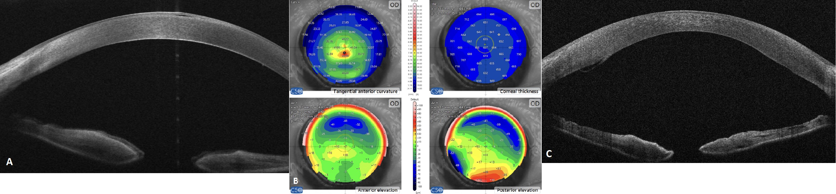
Fuchs' Endothelial Corneal Dystrophy (FECD) is a prevalent age-related eye disease affecting over 4.5% of individuals aged 50 and above. This condition is characterized by the progressive loss of corneal endothelial cells, leading to corneal oedema (swelling, Figure 1), pain, and vision impairment. Currently, the only definitive treatment available is corneal transplantation surgery. Anterior segment Optical Coherence Tomography (AS-OCT) is an advanced specialized imaging modality that provides high-resolution cross-sectional scans of the cornea, offering an unprecedented opportunity to detect early clinical signs of FECD and subtle post-transplant complications.
While recent advancements in artificial intelligence (AI) hold the potential to revolutionize ophthalmology, research on automated detection and grading of FECD remains limited. Hence, there is a unique opportunity to leverage deep learning models to enhance the diagnosis, prognostication, and management of patients with FECD.
Aim: Develop and validate automated tools to enhance management of FECD using medical images derived from AS-OCT.
As part of the UCL Institute of Ophthalmology's inherited eye disease research program, we have recruited the world's largest cohort of FECD patients from Moorfields Eye Hospital (the largest specialist eye centre in Europe). Moorfields Eye Hospital is equipped with the latest AS-OCT technology available in the United Kingdom. We have generated a vast AS-OCT imaging dataset that is exclusive to our research group (Figure 2). The student involved in this project will benefit from this unparalleled data resource for model training. Our research group has already conducted preliminary training of deep learning models on the existing dataset, yielding promising results. The outcomes of this project are likely to directly enhance patient care. This project will be under the guidance and supervision of Dr. Siyin Liu, Dr. Gunjan Naik, and Dr. Nikolas Pontikos.